A cone cell is a light-sensitive retinal photoreceptor that absorbs particular parts of the visible spectrum. The three types respond preferentially to short, medium, or long wavelengths, commonly associated with blue, green, and red. The brain blends their signals to produce color vision, a process called trichromacy. section 4.1.4 section 4.1.1
Cone-cell differences help explain several forms of color vision deficiency:
- Dichromacy occurs when one cone type is not functioning; protanopia, deuteranopia, and tritanopia correspond to absent red-, green-, or blue-associated responses. section 4.1.4
- Monochromacy occurs when two or all three cone types are not functioning; achromatopsia specifically describes complete inability to see color. section 4.1.4
- Anomaly occurs when all three cone types work but one has shifted or weaker sensitivity, as in protanomaly, deuteranomaly, or tritanomaly. section 4.1.4
A rainbow through cone cells
White sunlight contains many wavelengths. A prism separates them into a rainbow, and the cone cells absorb those wavelengths in different amounts, producing different color signals. section 4.1.1
Figure 4.8 This graph shows a normalized view of how much of each wavelength of visible light is absorbed by each type of cone in our eyes. S, M, and L refer to the short, medium, and long wavelengths of the light they absorb the most, also known as blue, green, and red, respectively, because that’s where the most light is absorbed. (BenRG, Public domain, via Wikimedia Commons)
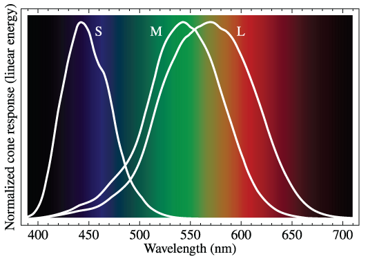
Use the normalized sensitivity graph to compare how short-, medium-, and long-wavelength cone cells absorb visible light. section 4.1.4