A pharmacophoric feature represents a molecular interaction capability: hydrogen-bond donation, hydrogen-bond acceptance, hydrophobicity, aromatic interaction, or metal-ion coordination. A pharmacophore combines these features with the geometry required for a compound to interact productively with a biological target. section 7.3.2
The representation separates what a molecule can do from its exact atom identities. Features may be annotated from a ligand alone or defined relative to a target structure, then compared across conformers to find recurring interaction patterns. section 7.3.2
A practical pharmacophore-modeling workflow proceeds as follows:
- Generate reasonable conformers for a ligand set. section 7.3.2
- Annotate each conformer with features such as donors, acceptors, and aromatics, including their three-dimensional coordinates. section 7.3.2
- Align conformers from different compounds to identify common features and measure their spacing. section 7.3.2
- Validate the resulting pattern on an external test set before using it to score screening compounds. section 7.3.2
From features to virtual screening
For a fragment ensemble, record donor, acceptor, and aromatic features, calculate distances between feature pairs, and fit kernel-density models to those distance distributions. Score a new molecule by how well its feature-pair distances fit the learned distributions, producing a structure-based virtual-screening signal. section 7.3.3
Figure 7.12 A) Morphine pharmacophore. B) Pharmacophore-based explanation for discrepancy in biological effects of adrenaline stereoisomers. C) Example pharmacophore features separated by a certain distance, which we can satisfy by selecting the shown functional groups and linking them to preserve desired spacing.
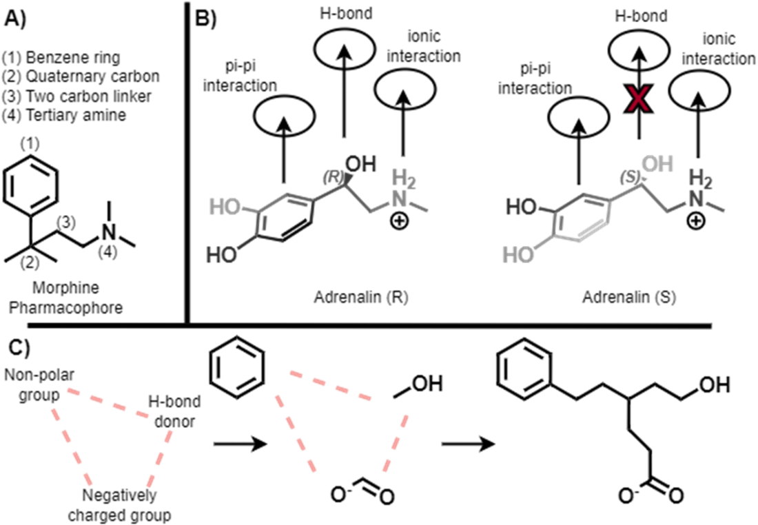
Use the figure to connect pharmacophore geometry with molecular activity: morphine illustrates a pharmacophore, adrenaline stereoisomers illustrate orientation, and linked groups illustrate spacing constraints. section 7.3.2