Structure preparation configures protein and ligand structures for reliable computational analysis, especially molecular docking. PDB structures may be incomplete or chemically ambiguous, so preparation decisions can change docking scores and rankings. section 9.1.3
A practical preparation checklist includes:
- Assign protonation states, tautomers, net charge, and partial charges at the relevant pH. section 9.1.3
- Model unresolved side chains, atoms, or loops near the binding site. section 9.1.3
- Decide whether to retain crystallographic waters, cofactors, and coordinated metals. section 9.1.3
- Preserve ligand chirality and double-bond geometry, then catch failed embedding, minimization, or atom typing. section 9.1.3
Extracting an EGFR receptor and ligand
For an EGFR PDB structure, load the file with MDTraj, select atoms matching "protein" for the receptor, select the ligand by residue name, and save separate PDB files. The EGFR construct has 41 unresolved residues, which may require modeling near the binding site. section 9.1.3
Figure 9.3 Wild type EGFR in complex with LN2057 obtained by X-ray diffraction (PDB ID: pdb_00006vhn). We represent EGFR as a Gaussian surface, which renders the 3D shape of the protein in a way that emphasizes its overall volume and shape (rather than a detailed atomic structure). We color EGFR by its accessible surface area, which renders the protein surface in colors ranging from white to dark blues. The least accessible (buried) residues are colored white. The darker blues reflect greater exposure of the corresponding residues. Refer to this video on Mol* for a tutorial on visualizing biomolecular structures using Mol* in PDB.
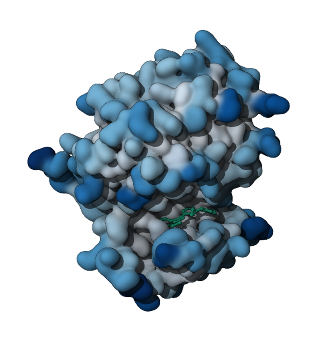
Visualize EGFR as a Gaussian surface colored by accessible surface area to inspect overall shape and residue exposure around the complex. section 9.1.3