A binding pose is a predicted three-dimensional position, orientation, and conformation of a ligand relative to a protein binding site. Docking searches possible poses, then ranks them with a scoring function that considers interactions such as van der Waals forces, electrostatics, hydrogen bonding, and desolvation. section 9.1.2
The preferred pose is the lowest-scoring configuration, treated as the most likely binding mode. Scores remain estimates: a pose can look geometrically plausible while still failing to predict potency, and confidence in geometry is not a potency measurement. section 9.1.2 section 13.2.2
Evaluate a predicted pose by checking:
- Affinity ranking, using the affinity column rather than minimizing every diagnostic energy component simultaneously. section 9.1.5
- Geometric plausibility, including favorable contacts, steric clashes, and unrealistic or strained ligand conformations. section 9.1.5 section 13.3.2
- Robustness, by comparing poses with a known structure, rescoring them, running molecular dynamics, or testing experimentally. section 9.1.5
Rentosertib in the TNIK ATP site
DiffDock’s rank-1 pose overlaid the rentosertib co-crystal structure at 0.92 Å symmetry-corrected heavy-atom RMSD and reproduced hinge contacts. Four runs produced 0.92, 0.81, 0.85, and 0.70 Å RMSD, but the result represents pose recovery rather than an unbiased blind prediction. section 13.2.2
Figure 9.2 Schematic overview of our protein-ligand docking workflow for structure-based drug design, illustrating a pipeline from obtaining the initial protein-ligand structure to analyzing molecular interactions in the docked complexes. The process consists of three main stages: (1) Input Preparation, where the EGFR-ligand complex structure is obtained from the PDB database and separated using MDTraj; (2) Structure Preparation, involving receptor preparation with PDBFixer, ligand preparation with Meeko, and binding site definition based on ligand coordinates; and (3) Docking & Analysis, where AutoDock Vina performs conformational sampling and pose evaluation, followed by post-processing and interaction analysis using ProLIF. Dark blue rectangles represent computational tools, while rectangles indicate processes within the docking engine, and light blue rectangles represent the data files we reference throughout this chapter’s Python notebook.
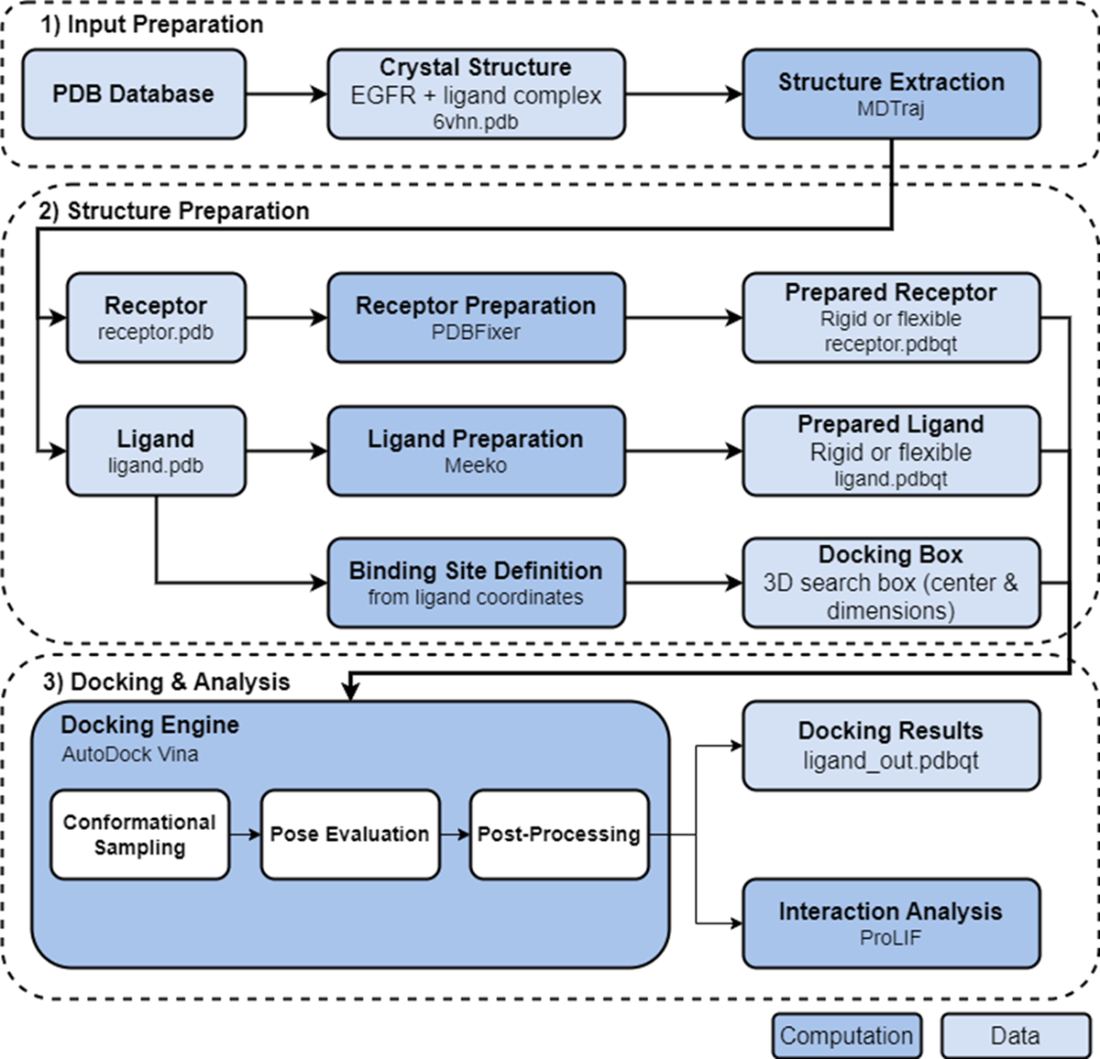
Follow the workflow from protein-ligand input preparation through receptor and ligand preparation, docking, pose evaluation, and interaction analysis. section 9.1.2