A Hounsfield unit (HU) measures x-ray radiodensity in computed tomography (CT). Each voxel in a CT reconstruction receives a value estimating how strongly its material absorbs x-ray radiation, using water as the reference point. section 12.1
Useful reference values make HU practical for interpreting CT data:
- Air is approximately −1000 HU, while water is exactly 0 HU. section 12.1
- Soft tissue is about 100 HU; bone commonly ranges from roughly 300 to 2000 HU. section 12.1
- Metal can reach values as high as 30,000 HU. section 12.1
CT scanners collect x-ray measurements from multiple angles. A computer combines the differing energy losses by solving linear systems, producing a 3D image whose voxels contain HU values. section 12.1
Selecting bone with an HU threshold
To isolate likely bone from a CT volume, keep voxels between 300 and 3000 HU and discard the rest. This simple segmentation rule removes much of the surrounding fat, muscle, and air before surface modeling or 3D printing. section 12.2
Thresholds are useful but approximate. Bone marrow is less dense than cortical bone, joint regions can be porous, and CT may not represent porosity accurately. A threshold can therefore be too narrow or too broad, requiring later surface cleanup. section 12.2
Figure 12.2 A CT scan scheme (FDA, public domain, via Wikimedia Commons), and our simplified example. We take two 1D x-ray pictures at a different angle (first b11, b12 and then b21, b22) to compute a single 2D scan ([[a11, a12], [a21, a22]]).
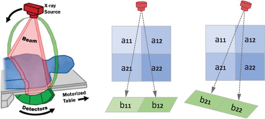
Use the simplified CT diagram to connect multiple angled x-ray measurements with the reconstructed 2D array of radiodensity values. section 12.1