Radiodensity measures how strongly a material absorbs X-rays. In computed tomography (CT), each voxel stores an estimated radiodensity value, allowing many 2D scans to form a 3D image. section 12.1
Hounsfield units (HU) provide a practical scale:
- Air: about −1000 HU; water: exactly 0 HU. section 12.1
- Soft tissue: about 100 HU; bone: roughly 300–2000 HU. section 12.1
- Metal can reach approximately 30,000 HU. section 12.1
A scanner compares emitted and detected X-ray energy. Each ray records the combined absorption along its path; measurements from many angles support reconstruction by solving linear systems. section 12.1
Isolating bone with a threshold
A simple segmentation rule keeps voxels from 300 to 3000 HU and filters out much surrounding tissue. Narrow thresholds miss less-dense bone, while broad thresholds admit noise. section 12.2
Figure 12.2 A CT scan scheme (FDA, public domain, via Wikimedia Commons), and our simplified example. We take two 1D x-ray pictures at a different angle (first b11, b12 and then b21, b22) to compute a single 2D scan ([[a11, a12], [a21, a22]]).
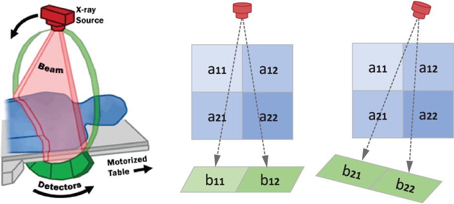
Use the CT scheme to connect two angled one-dimensional X-ray measurements with reconstruction of a small two-dimensional radiodensity grid. section 12.1
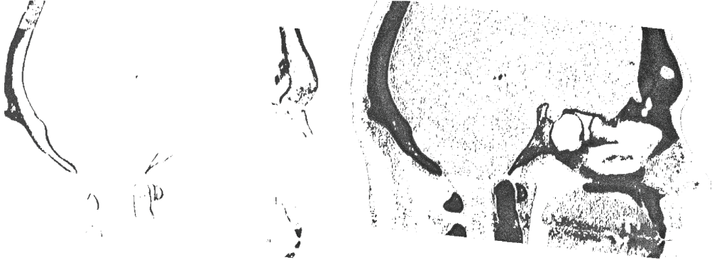
Compare thin and broad thresholds to see the practical balance between missing bone and retaining noise. section 12.2