DiffDock predicts how a small molecule, or ligand, may fit into a protein and assigns confidence scores to the predicted binding poses. It uses a diffusion model trained on experimental protein–ligand complexes: starting from a random ligand placement near the protein, it iteratively refines the placement into a plausible bound geometry. section 13.2.1 section 13.3.2
Pose confidence is a geometric signal, not a binding-affinity or potency estimate. A confident pose can suggest how a ligand might bind, but it does not establish how strongly the ligand binds or whether it will work experimentally. section 13.2.1 section 13.2.2
Use DiffDock as one stage in a screening workflow:
- Provide a protein structure and candidate ligands; generate and rank predicted poses with their confidence scores. section 13.3.2 section 13.2.1
- Check pose geometry as well as confidence. Discard poses with protein-atom clashes or strained, high-energy ligand conformations. section 13.3.2
- Evaluate surviving candidates for properties such as solubility and toxicity, then use expert review and experiments to validate them. section 13.3.2 section 13.5
On the original PDBBind blind-docking benchmark, roughly 38% of DiffDock’s top-ranked poses were within 2 Å of the crystallographic pose, compared with about 23% for traditional search-based docking and 20% for the previous best deep-learning method. The distance measure, RMSD, summarizes how far predicted atoms are from their reference positions. These are benchmark averages, not guarantees for an unfamiliar target or binding mode. section 13.2.1
TNIK pose recovery
For rentosertib bound to TNIK, the top-ranked pose was 0.92 Å symmetry-corrected heavy-atom RMSD from the co-crystal pose and was the best of 20 sampled poses. Four independent runs returned 0.92, 0.81, 0.85, and 0.70 Å. This is pose recovery, not a clean blind test: the co-crystal is in the PDB, and the served weights use PDB-derived data. Across 18 TNIK ligands, rank-1 confidence did not track measured potency: Spearman correlation was −0.245. section 13.2.2
Figure 13.5 The rank-1 DiffDock pose of rentosertib overlaid on its crystallographic pose in the TNIK ATP site (PDB 8ZML), at 0.92 Ångströms symmetry-corrected heavy-atom RMSD. The hinge contacts that define the binding mode, the Met105 gatekeeper and the Cys108 hinge residue, are reproduced, and the rank-1 pose is also the best of the 20 sampled, so the confidence ranking selected the right one rather than merely including it. Four independent runs returned 0.92, 0.81, 0.85, and 0.70 Å. Note this is pose recovery rather than blind prediction, as 8ZML is a rentosertib co-crystal in the PDB and the served weights are trained on PDB-derived data, so the complex may sit inside the model's training distribution.
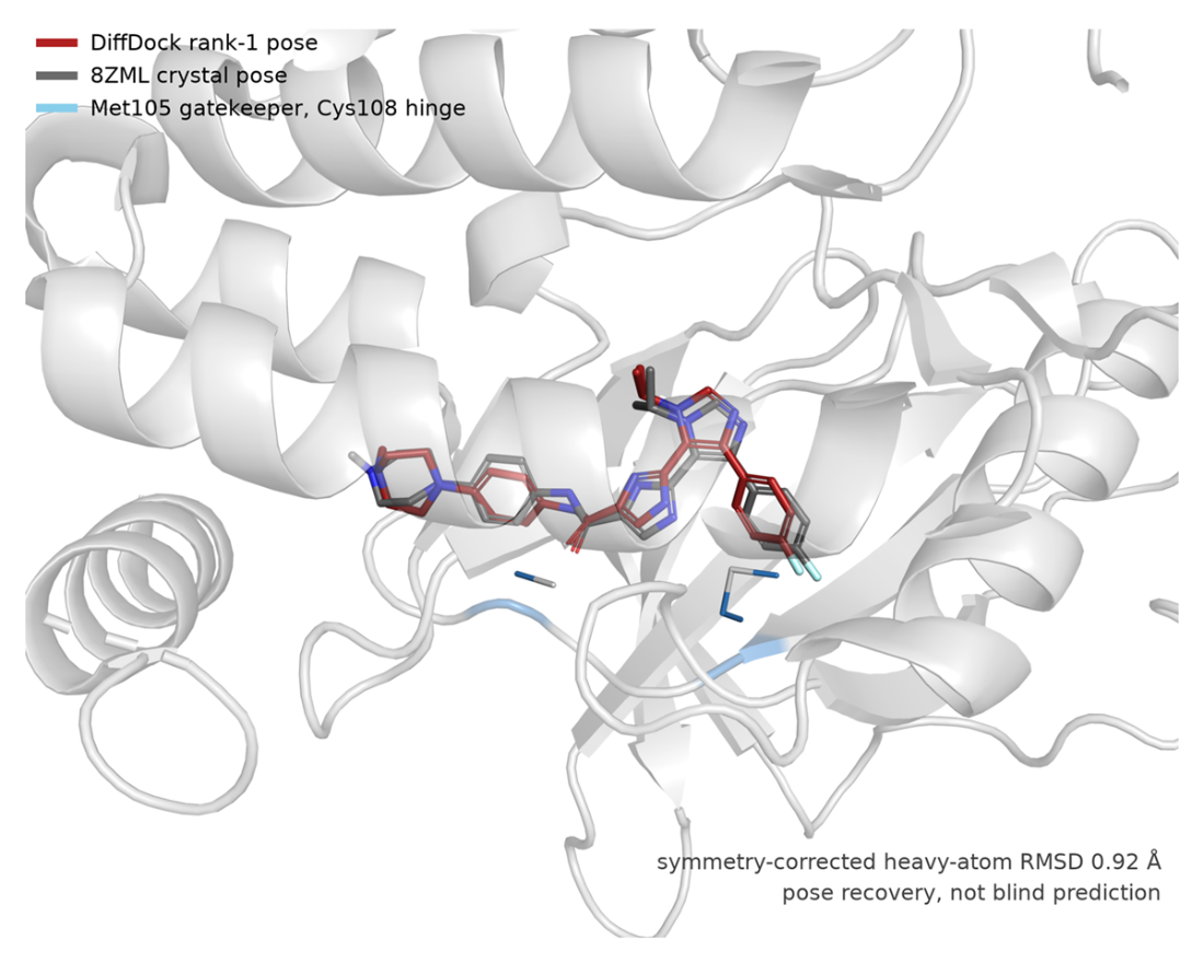
Compare the top-ranked rentosertib pose with its crystallographic reference and inspect the reproduced binding-site contacts. section 13.2.2
Filtering a large library
In a FADS1 screening workflow, an embedding-based filter reduced 4.3 billion compounds to 10 million before DiffDock screened them against 20 protein structural variants. Confidence and geometric filters reduced the set to 50,000 for property screening. Screening at this scale requires substantial infrastructure and downstream experimental validation. section 13.3.2 section 13.2.1