Computed tomography (CT) reconstructs internal structures from many x-ray measurements taken around an object. A scanner compares emitted radiation with the lower energy detected after passage through the body, then combines measurements from different angles into a 3D image whose voxels store radiodensity. section 12.1
The reconstruction process connects physical measurements with a small mathematical model:
- A single x-ray view records total radiodensity along each ray, but cannot uniquely locate individual bones, tissues, or airways. section 12.1
- Many views provide enough constraints to compute the unknown voxel values by solving a series of linear systems. section 12.1
- A four-pixel 2D slice makes the geometry concrete: two two-pixel x-ray scans from different angles help determine values a11, a12, a21, and a22. section 12.1
From a scanned part to a replacement
CT can capture a train, ship, or airplane part whose original blueprints are unavailable. The scan can support a boundary representation, which can then be milled, converted into a triangle mesh for 3D printing, or retained as smooth parametric surfaces for manufacturing. section 12.5
Figure 12.2 A CT scan scheme (FDA, public domain, via Wikimedia Commons), and our simplified example. We take two 1D x-ray pictures at a different angle (first b11, b12 and then b21, b22) to compute a single 2D scan ([[a11, a12], [a21, a22]]).
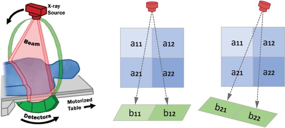
Use the scan scheme to relate angled 1D x-ray measurements to the reconstructed 2D pixel grid. section 12.1